Effect of Crystallographic Artifacts for Structure Prediction and Design
Published
Cite this note:Ramith Hettiarachchi (2026, November 16). Effect of Crystallographic Artifacts for Structure Prediction and Design. ramith.fyi. https://ramith.fyi/notes/2026-11-16-effect-of-crystallographic-artifacts-for-structure-prediction-and-design/.
I was reading this paper [1] by El Nesr et al. in which they highlight a very interesting fact on how crystallographic artifacts in the PDB can bias structure prediction models and design models.
El Nesr et al. introduce dEVA to design de novo zinc-binding proteins and metalloenzymes. dEVA is an evolutionary design approach that alternatively refines 1) sequence + side-chain proposals from LigandMPNN [2] with 2) metal-site predictions from Metal3D [3] on a fixed starting backbone.
In this post we will be focusing on a particular negative control (desD1) that they tested in wet lab experiments.
After the dEVA design process, its binding site had only a single coordinating residue (Asp63) within binding distance while Glu65 pointed into solvent.
Now, how would AF3 predict this? Surprisingly similarly: Glu65 still points to the solvent and the metal ion is interacting with Asp63. So in the standard of “refolding” similarity, this is a refolding success11 it predicted the metal ion in that exact spot with high confidence (Zn pLDDT: 86.55).
However, when they tested this negative control experimentally, they found that it’s an incomplete coordination environment that showed no detectable Zn2+ binding22 neither by MagFura-2 competition titration nor by ICP-MS.
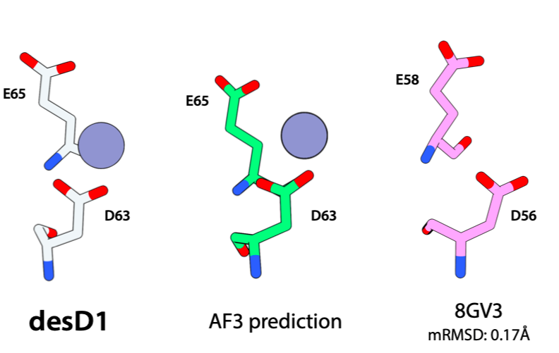
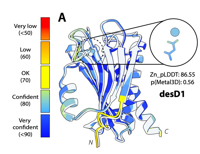
desD1 came from an unconditional Protpardelle backbone packed with metal-blind LigandMPNN, onto which Metal3D placed a low-confidence putative Zn2+ (p = 0.56) coordinated only by D63; AF3 reproduces that single-residue pose (green), which geometrically matches an ordinary non-metal-binding surface turn (D56/E58 in PDB: 8GV3, 0.17 Å mRMSD). Right (Fig. 3A): AF3 fold of desD1 colored by pLDDT overlaid on the design (Zn_pLDDT: 86.55). Adapted from El Nesr et al. [1] (CC BY 4.0).Rerunning the AF3 prediction
To analyze this example further, I folded the sequence of desD1 with AF3 server paired with a single ZN CCD ligand token:
>desD1 | dEVA negative control (177 aa; p(seq)=0.48, p(metal)=0.56)
AMLPASDFFEETVPGTVTFKEVVADPESPGSQKVLDRLIDLALDAASKVPGRIVTFIFRF
RQDPEDPNGWQIDILIYVDGKPVAVAGVTFNTDGTLTFYRRGITAEEYDALEALPAILRI
TIVVDVEAAKILEREVEVSPLVSELGLSKEEALASAEIIADFATKKLVAWARELRAA
{
"name": "desD1_zn",
"modelSeeds": [1],
"sequences": [
{ "protein": { "id": "A", "sequence": "AMLPASDFFEETVPGTVTFK..." } },
{ "ligand": { "id": "B", "ccdCodes": ["ZN"] } }
],
"dialect": "alphafold3",
"version": 1
}Figures below show the AF3 co-folding results:
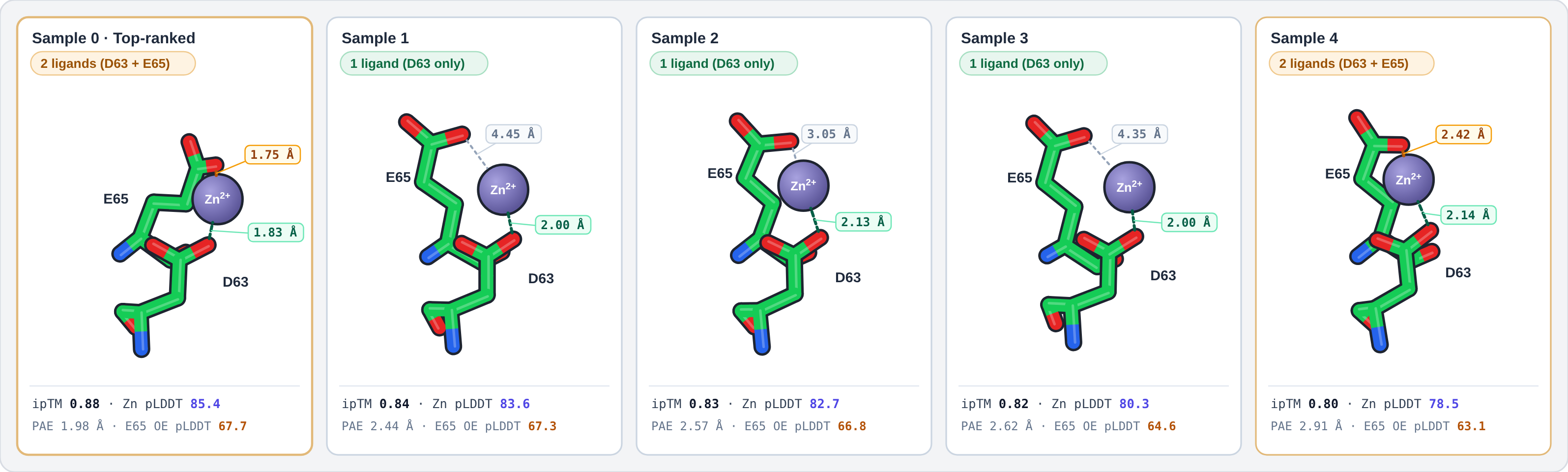
seed = 373020228, Sample 0–Sample 4) for desD1 + 1 × ZN, superposed on the Asp63–Pro64–Glu65 β-hairpin (N, Cα, C of residues 61–67) and viewed from the same orientation as Fig. S5. Dashed green/amber lines mark O–Zn contacts ≤ 3.0 Å; grey dotted lines mark non-coordinating Glu65 distances into solvent. Hover either mode here (or any card above) to compare 1-ligand (Asp63 only) vs. 2-ligand (Asp63 + Glu65) rollouts.to be continued.
References
- El Nesr et al. (2026). “Zero-shot design of a de novo metalloenzyme.”
- Dauparas et al. (2025). “Atomic context-conditioned protein sequence design using LigandMPNN.”
- Dürr et al. (2023). “Metal3D: a general deep learning framework for accurate metal ion location prediction in proteins.”
MoveThanks!